Ding, Z., Fahey, P. G., Papadopoulos, S., Wang, E. Y., Celii, B., Papadopoulos, C., … & Tolias, A. S. (2025). Functional connectomics reveals general wiring rule in mouse visual cortex. Nature, 640(8058), 459-469. https://www.nature.com/articles/s41586-025-08840-3
In vivo calcium imaging combined with EM connectomics
Mouse visual cortex
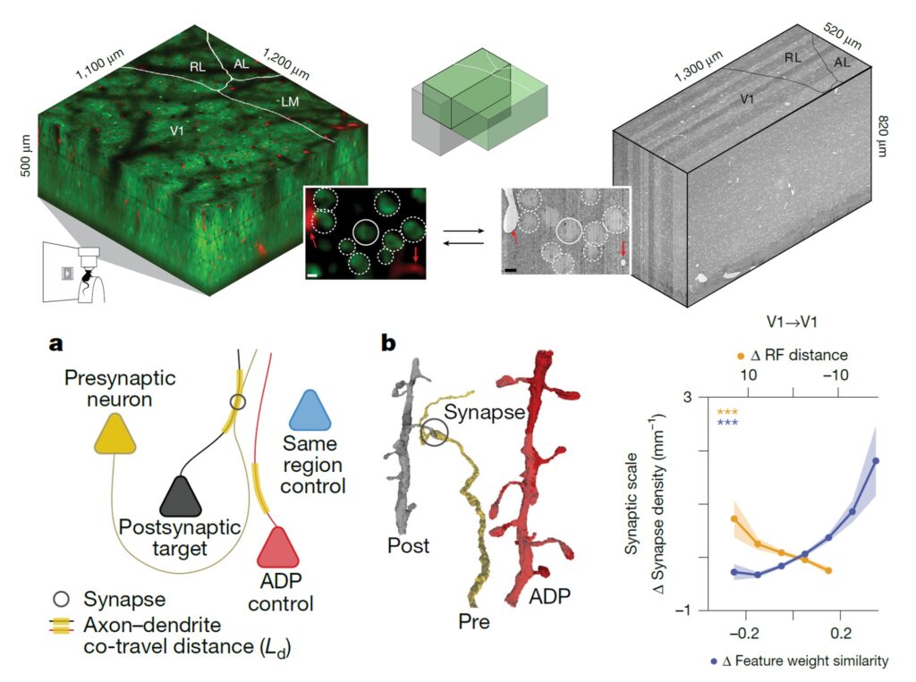
This paper summarizes some of the key findings of the MICrONS project -perhaps the most ambitious connectomics project to date. The MICrONS project acquired a cubic millimeter volume electron microscopy dataset spanning mouse primary and higher-order visual areas. Overlapping regions were calcium imaged in the awake behaving mouse while watching hours of specially designed video stimuli. This paper analyses a subset of the neurons having both high-quality calcium recordings and proofread connectome tracings. This allowed the authors to ask questions like “Are neurons that respond to similar visual features more likely to be synaptically connected?” The authors employ two novel analysis techniques: 1.) They quantify the distance each axon travels in close proximity to each dendrite. This allows them to normalize the actual synapse density seen between any two cells. 2.) The authors employ a ‘digital twin’ to precisely quantify each cell’s receptive field location tuning and feature tuning separately. Using these, the authors show that cells with similar feature tuning have higher than expected synaptic interconnectivity. Intriguingly they show that cells with similar location tuning show lower than expected synaptic interconnectivity. Results that have significant implications for our models of learning in cortex.
“Using a validated digital twin model, we separated neuronal tuning into feature (what neurons respond to) and spatial (receptive field location) components. We found that only the feature component predicts fine-scale synaptic connections beyond what could be explained by the proximity of axons and dendrites.”